13,276 anatomy of the head and neck stock photos, vectors, and illustrations are available royaltyfree See anatomy of the head and neck stock video clips of 133 muscle head anatomy vocal organ diagram female neck anatomy neck wireframe head neck human anatomy head artery anatomy face pharynx vector neck degree head anatomy 3dPalatine (2) – situated at the rear of oral cavity and forms part of the hard palate Maxilla (2) – comprises part of the upper jaw and hard palate Vomer – forms the posterior aspect of the nasal septum Mandible (jaw) – articulates with the base of the cranium at the temporomandibular joint (TMJ)The occipital bone is the trapezoidshaped bone at the lowerback of the cranium (skull) The occipital bone houses the back part of the brain and is one of seven bones that come together to form the skull It is located next to five of the cranium bones
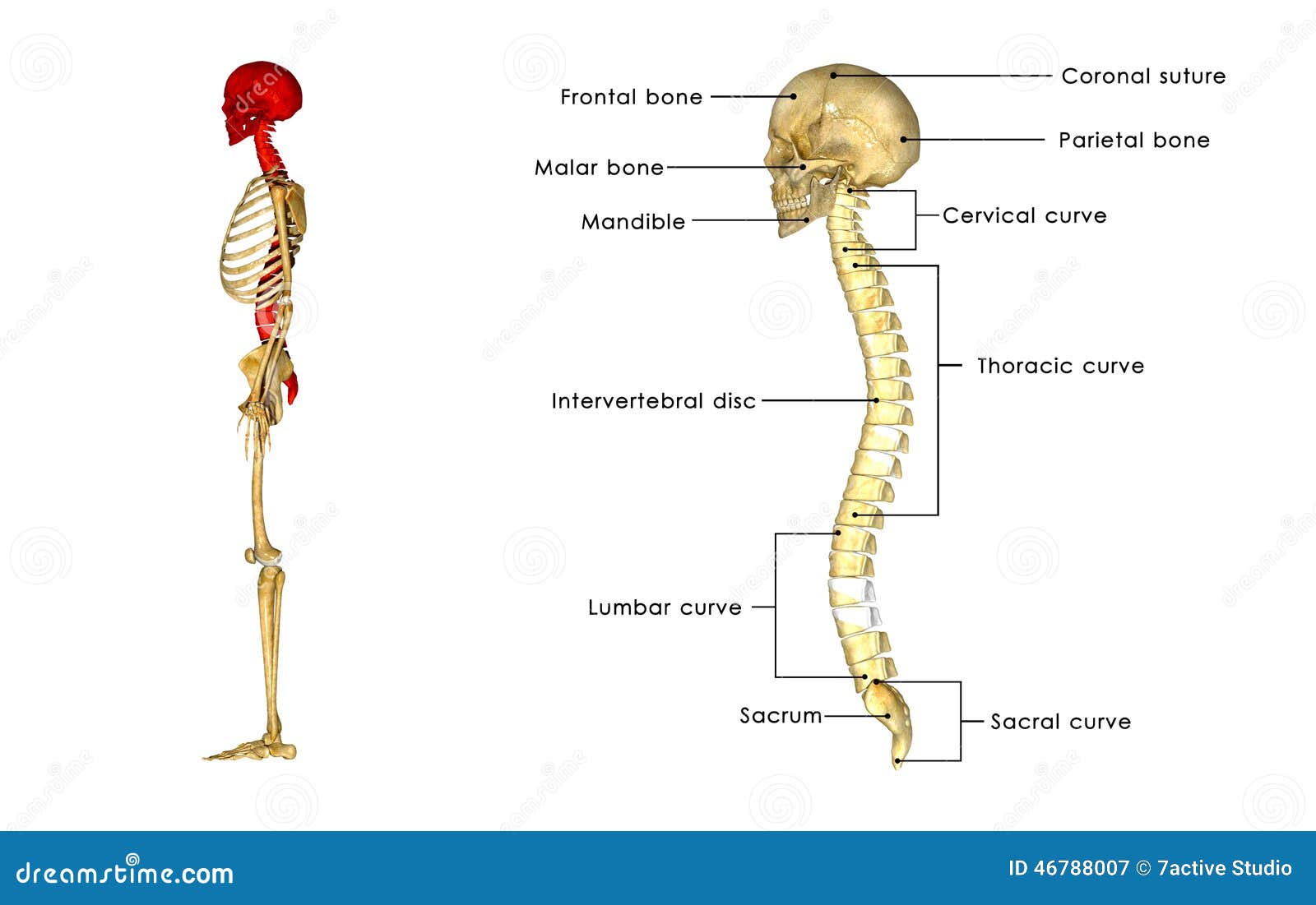
Back Bone With Skull Side View Stock Illustration Illustration Of Head Gland
Back of head bone anatomy labeled
Back of head bone anatomy labeled-6 rowsKey facts about head and neck anatomy Skull Comprised of 22 bones (Frontal bone, parietalHead hed 1 the anterior or superior part of a structure or organism 2 in vertebrates, the part of the body containing the brain and the organs of special sense Called also caput articular head an eminence on a bone by which it articulates with another bone head injury traumatic injury to the head resulting from a fall or violent blow Such an
:background_color(FFFFFF):format(jpeg)/images/library/9490/skull-posterior-lateral-views_english.jpg)


Posterior And Lateral Views Of The Skull Anatomy Kenhub
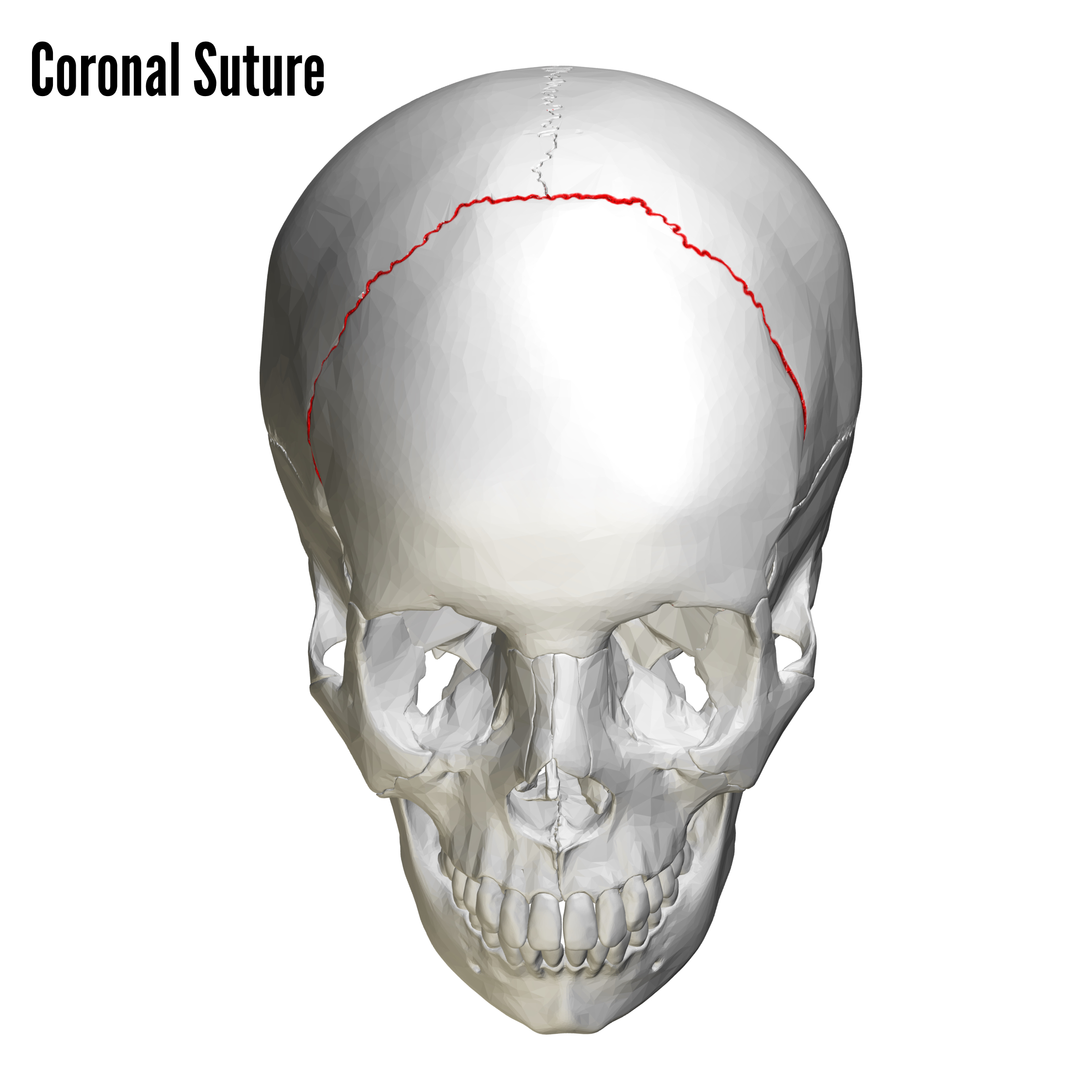
Skull anatomy Frontal Bone Parietal Bone Sagital Suture Coronal Suture the large cranial bone forming the front part of the cranium either of two skull bones between the frontal and occipital bo Located on the midline of the skull lies between both parietal the suture between the parietal and frontal bones of the skullLearn more about the hardest working muscle in the body with this quick guide to the anatomy of the heart The spine is the backbone of the human skeleton The upper arm or brachium, which is the region according to healthline, the human arm is composed of three bones, divided amongst tw Human Bone Anatomy Back It is also flexible enough toThe cranium can be divided further in to the calvarium and the cranial base The calvarium is comprised of the frontal, occipital and two parietal bones, and the cranial base is comprised of the frontal, sphenoid, ethmoid, occipital, parietal and temporal bones
Back Of Neck Anatomy Bones If you can, buy or borrow a we At the elbow, it connects primarily to the ulna, as the forearm's radial bone connects to the wrist The humerus is the long bone in the upper arm It sits on the radial or lateral side of the wrist near the thumb It is made up of 24 bones known as vertebrae, according to spine universeThe cervical spine supports the weight and movement of your head and protects the nerves exiting your brain The lumbar spine – the lower back, composed of five vertebrae, provides support for the majority of your body's weight The thoracic spine – the middle back, made up of the 12 vertebrae in between the cervical and lumbar spineThe semispinalis capitis, splenius capitis, and longissimus capitis muscles all help the head extend toward the back They also work with sternocleidomastoid muscles to rotate the head left and right
Occipital condyle where the skull articulates with the atlas (first cervical vertebrae) Foramen lacerum Fibrocartilagefilled hole between the body of the sphenoid bone and the petrous part of the temporal bone Digastric notch the posterior belly of the digastric muscle attaches to this pit Mastoid processThe muscle has a frontal belly and an occipital (near the occipital bone on the posterior part of the skull) belly In other words, there is a muscle on the forehead ( frontalis) and one on the back of the head ( occipitalis ), but there is no muscle across the top of the headThe human skull (Latin cranium) is the skeleton of the head composed of 22 separate bones joined together primarily by suturesMost of the bones have pairs The skull by Anatomy Next The brain is located within the skull and connected with other anatomical structures by the nerves and blood vessels going through many foramina (openings), and the largest foramen of the skull
:watermark(/images/watermark_only.png,0,0,0):watermark(/images/logo_url.png,-10,-10,0):format(jpeg)/images/anatomy_term/skull/eEsfu70EOMx1TlBf5tYAiA_Go0bFvBvzClwSivuaiELg_head_01.png)


Bones Of The Head Skull Anatomy Kenhub


Upper Cervical Spine Disorders Anatomy Of The Head And Upper Neck
Head and neck anatomy is important when considering pathology affecting the same area In radiology, the 'head and neck' refers to all the anatomical structures in this region excluding the central nervous system, that is, the brain and spinal cord and their associated vascular structures and encasing membranes ie the meningesCalcaneus The calcaneus (Latin calcaneus, calcaneum, meaning "heel") is the sizeable bone forming the heel It is the largest bone of the foot The calcaneus articulates with both the cuboid bone and talus within the tarsus The calcaneus acts as a short lever for the calf muscles, which insert into its posterior surface via the AchillesThe Crown begins at the point where the top of the head begins to curve downward to the back of the head and ends at the point just above the Occipital bone It is a semicircular area Occipital BoneThe Occipital Bone is the small bony protrusion at


Back Bone With Skull Side View Stock Illustration Illustration Of Head Gland
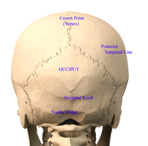


Back Of Head Skull Anatomy Dr Barry Eppley Indianapolis Explore Plastic Surgery
The muscle has a frontal belly and an occipital (near the occipital bone on the posterior part of the skull) belly In other words, there is a muscle on the forehead ( frontalis) and one on the back of the head ( occipitalis ), but there is no muscle across the top of the head50,975 human back anatomy stock photos, vectors, and illustrations are available royaltyfree See human back anatomy stock video clips of 510 human body anatomy female female anatomy muscle shoulder blade pain anatomy back muscles bones man female anatomy body muscles in a body female anatomy muscole shoulder concept muscular sysyemThe first cervical vertebra (atlas) supports and balances the head The second vertebra (axis) allows the head to rotate laterally to the left and the right Hollow spaces within the cervical vertebrae protect and conduct the spinal cord and vertebral arteries through the neck
/male-skull-in-profile-with-transparent-head-on-white-background-1092338382-d031fe3a88fa4462b0ba082a1ec64302.jpg)


Occipital Bone Anatomy Function And Treatment
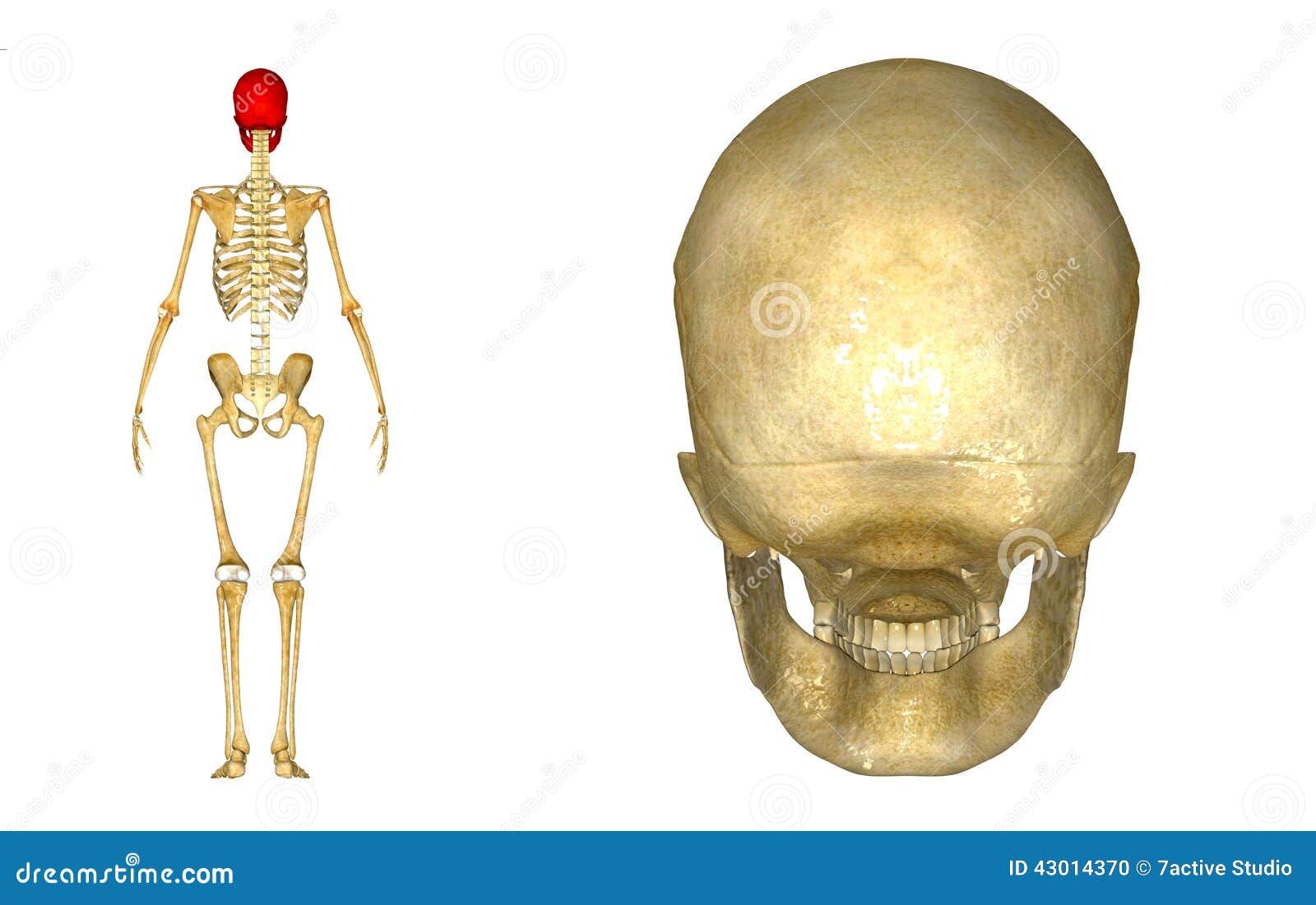


Human Skull Back Stock Illustration Illustration Of Graveyard
The collar bone connects with the acromion of the shoulder blade Other parts of the shoulder blade you must know are the coracoid process and the glenoid fossa (which is part of the shoulder joint) The head of humerus, or upper arm bone, is another part of the shoulder joint The full lesson is avaibale to Anatomy Master Class membersGross Anatomy of Bone The structure of a long bone allows for the best visualization of all of the parts of a bone ()A long bone has two parts the diaphysis and the epiphysisThe diaphysis is the tubular shaft that runs between the proximal and distal ends of the boneThe joint of head of rib (costocorporeal joint) is the articulation between the rib head and
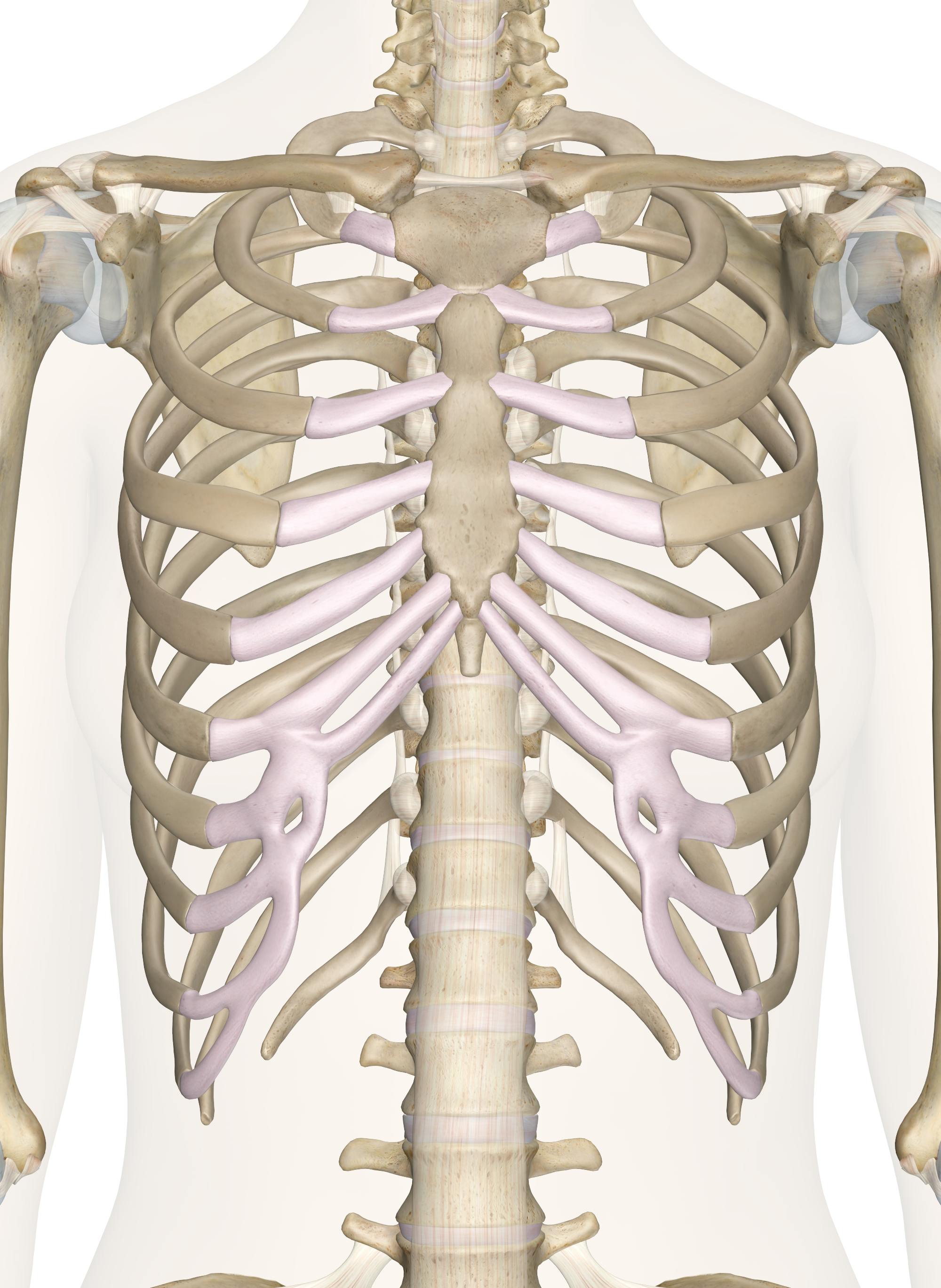


Bones Of The Chest And Upper Back
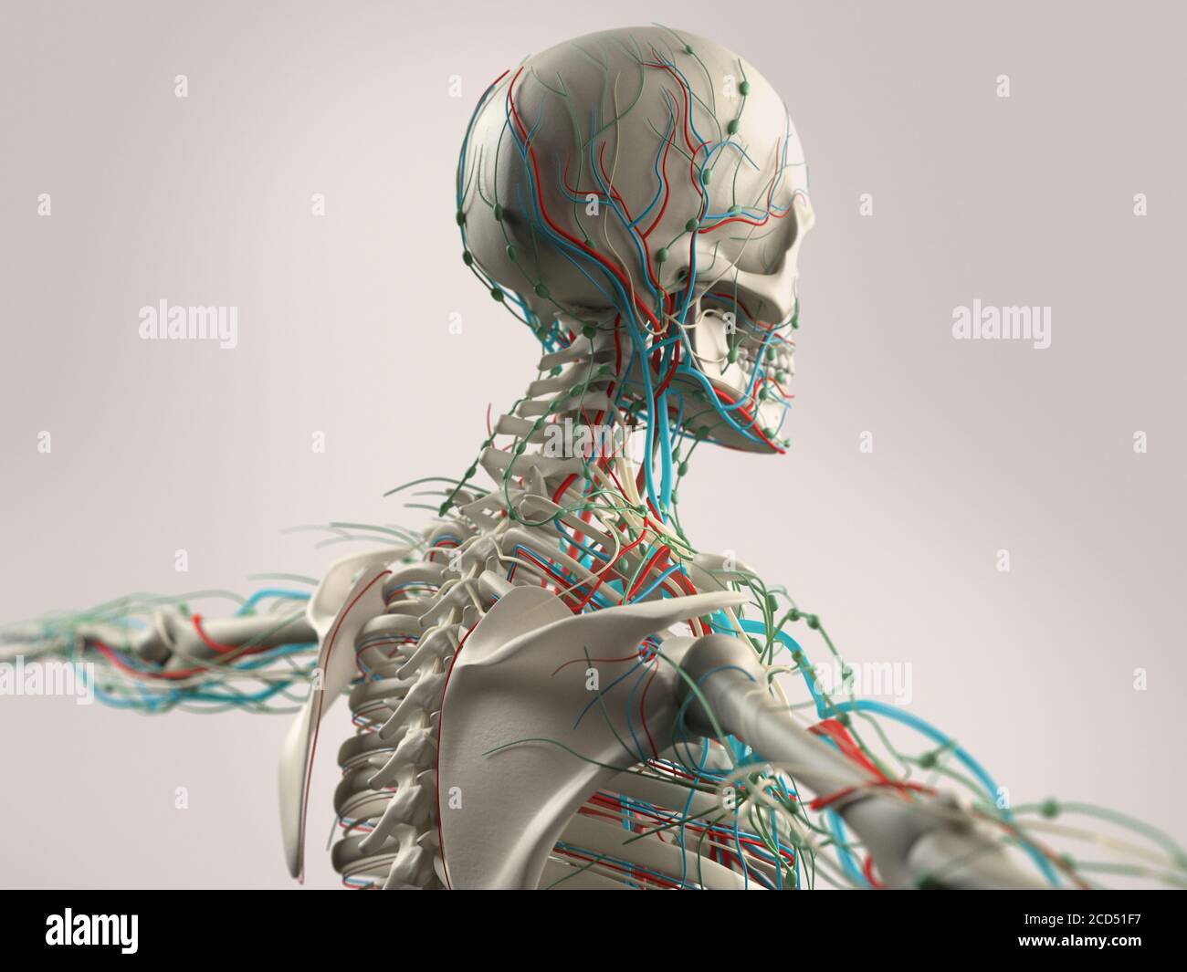


Human Anatomy Showing Face Head Shoulders And Back Muscular System Bone Structure And Vascular System Stock Photo Alamy
Browse 3,133 anatomy of neck and shoulder stock photos and images available, or start a new search to explore more stock photos and images Pain in a man's body Pain in a man's body on a gray background Collage of several photos with red dots anatomy of neck and shoulder stock pictures, royaltyfree photos &
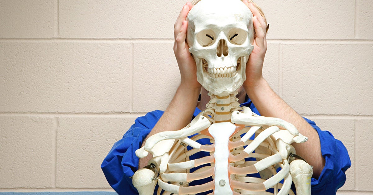


Flat Bones Definition Examples Diagram And Structure
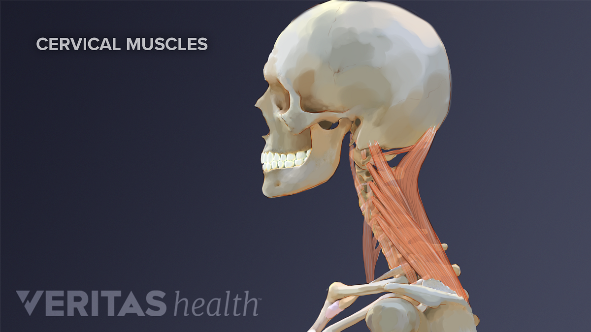


Neck Muscles And Other Soft Tissues
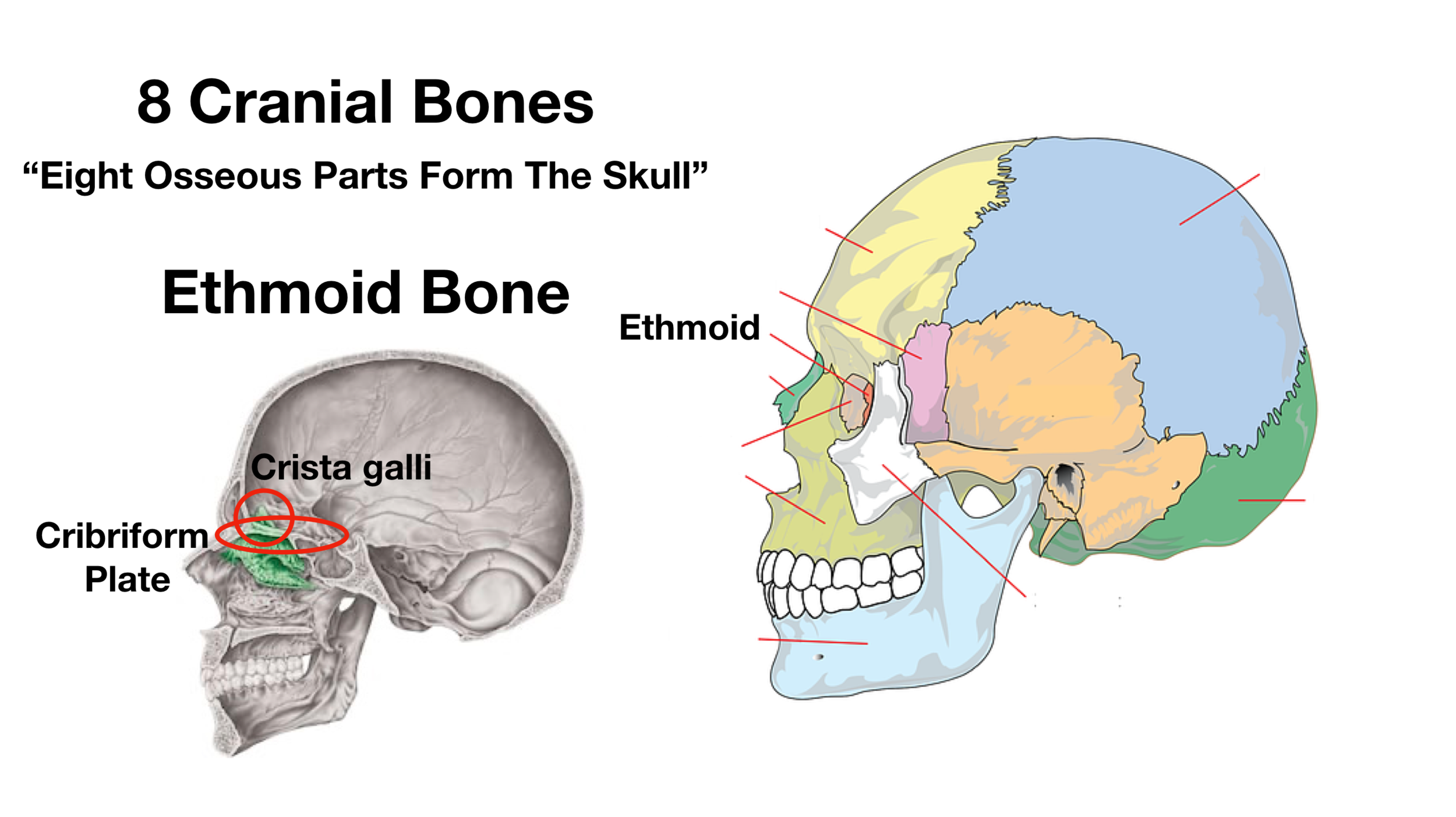


Skull Anatomy Cranial Bone And Suture Labeled Diagram Names Mnemonic Ezmed
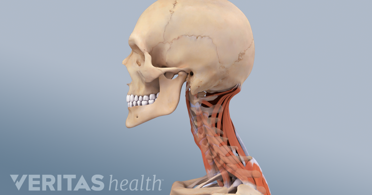


Cervicogenic Headache Causes And Risk Factors


Cranial Osteopathy Knowledge Amboss


Bone Structure Of The Face An Overview Of Dental Anatomy Continuing Education Course Dentalcare Com


Human Head Skull And Cervical Vertebrae Rear View Stock Photo Download Image Now Istock


Occipital Bone Wikipedia


Very Detailed And Scientifically Correct Human Skull Back View On White Background Anatomy Image Stock Photo Picture And Royalty Free Image Image


Bones Of The Head Atlas Of Anatomy


Baby Skeleton How Many Bones Are Babies Born With Babycenter


Occipital Bun Wikipedia


Anatomy Back Of Skull 2 Diagram Quizlet


Skull Anatomy Anatomy Bones Human Skull Anatomy


Occipital Ridge Wikipedia


3
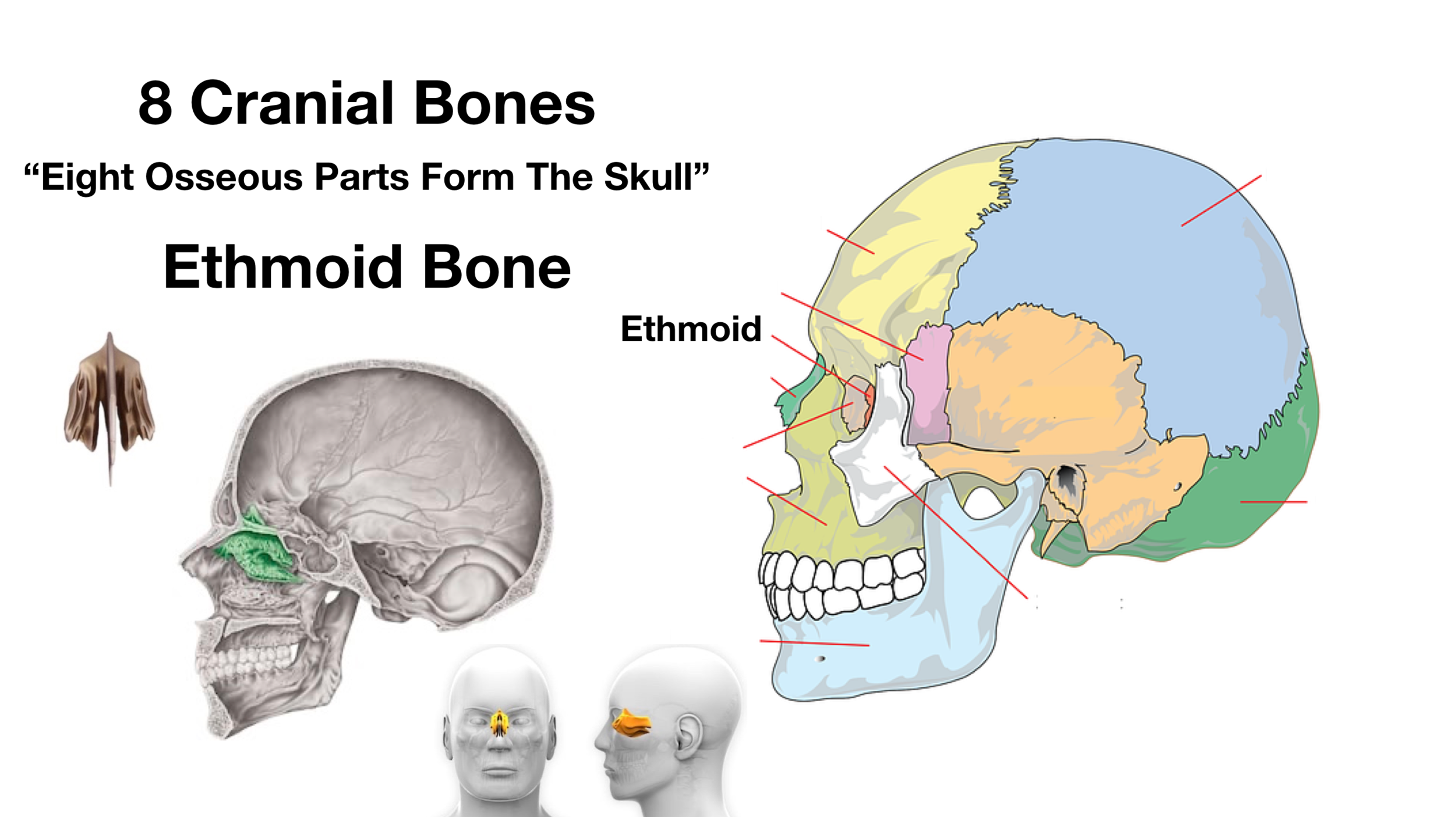


Skull Anatomy Cranial Bone And Suture Labeled Diagram Names Mnemonic Ezmed


Skull Anatomy Terminology Dr Barry L Eppley
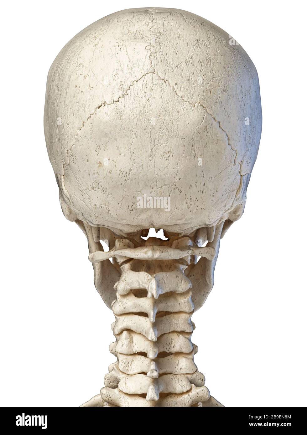


Medical Human Head Illustrations High Resolution Stock Photography And Images Alamy
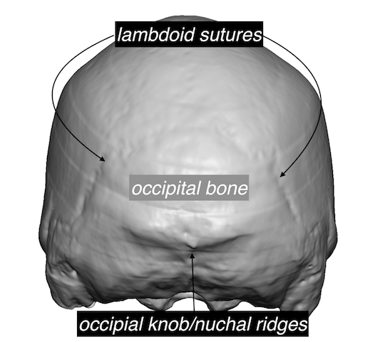


Plastic Surgery Case Study Occipital Skull Reduction With Parietal Onlay Bone Grafting For Back Of Head Reshaping Explore Plastic Surgery


Skull Functions Facts Fractures Protection View Bones
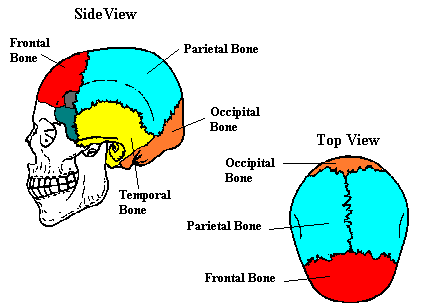


Neuroscience For Kids The Skull


Bones Of The Head And Neck Skull And Cervical Spine Preview Human Anatomy Kenhub Youtube


12 Types Of Bump On The Back Of The Head


Human Body Skull Anatomy External Occipital Protuberance Human Back Of Skull Face Human Png Pngegg


External Occipital Protuberance Wikipedia
:background_color(FFFFFF):format(jpeg)/images/article/en/posterior-and-lateral-views-of-the-skull/U0Gu2npm5ZRSP1eZgc4Jbw_Posterior_view_of_skull.png)


Posterior And Lateral Views Of The Skull Anatomy Kenhub
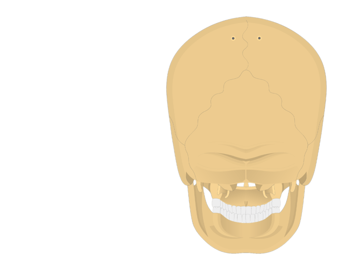


Occipital Bone Anatomy


1


Back View Of Bone Structure In The Head Stock Photos Page 1 Masterfile
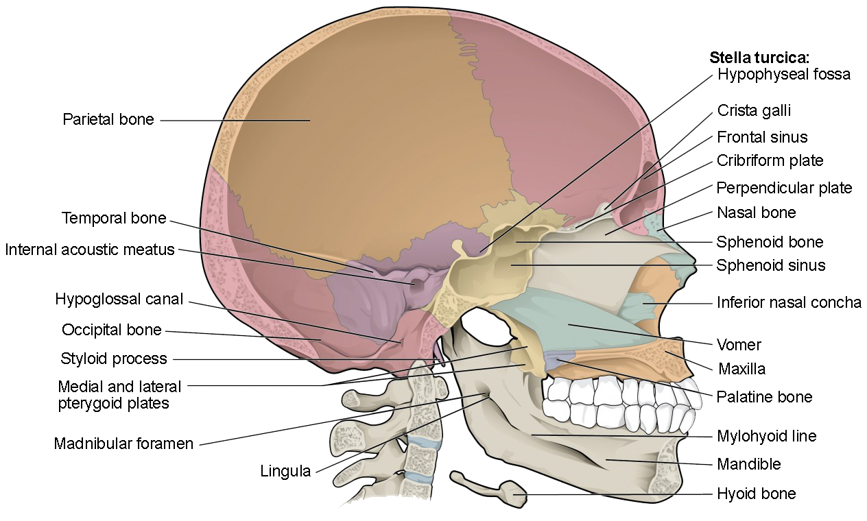


The Skull Anatomy And Physiology I
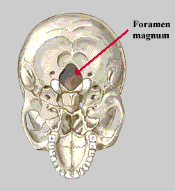


Neuroscience For Kids The Skull


Poor Posture Due To Smartphone Use Leads To Horn Bone Growth In Skull Youtube


The Skull Anatomy And Physiology I
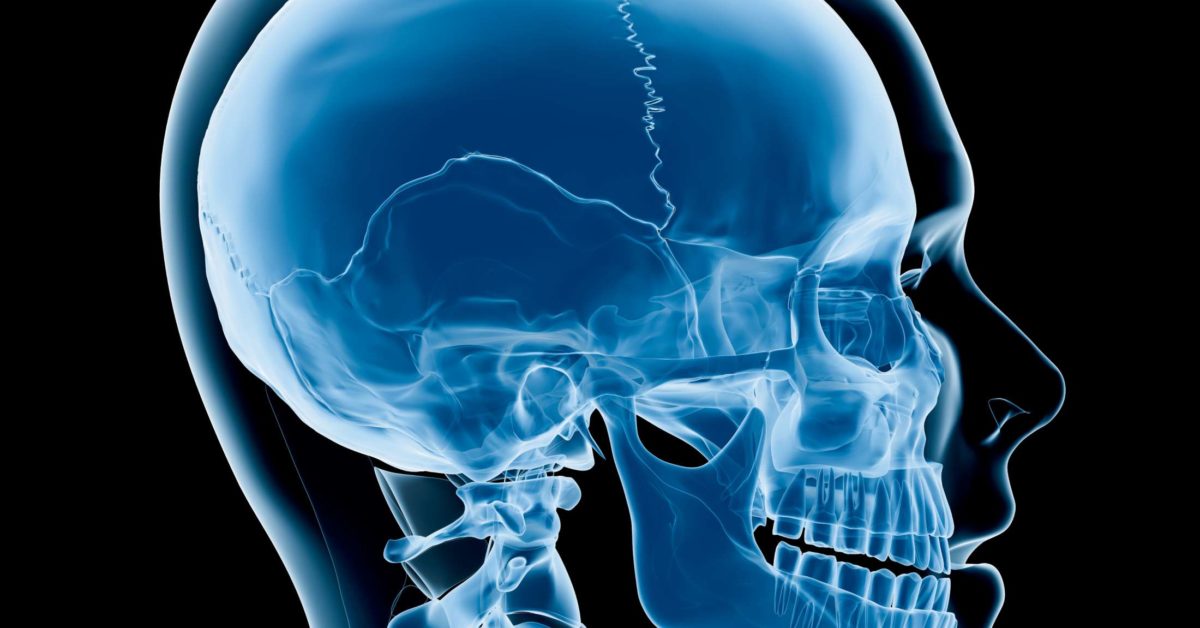


Craniosynostosis Types Causes Diagnosis And Treatment
:watermark(/images/watermark_only.png,0,0,0):watermark(/images/logo_url.png,-10,-10,0):format(jpeg)/images/anatomy_term/coronal-suture/VkKPXrZ4iRLzKUenK4ZVYg_Coronal_suture_02.png)


Skull Joints And Sutures Anatomy And Functions Kenhub


1
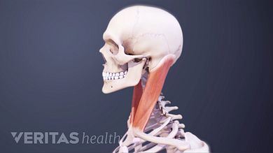


Neck Muscles And Other Soft Tissues
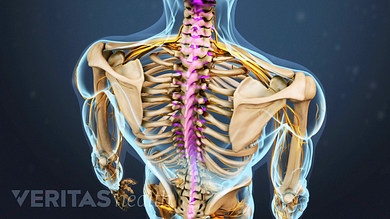


Spinal Anatomy And Back Pain


The Skull Boundless Anatomy And Physiology
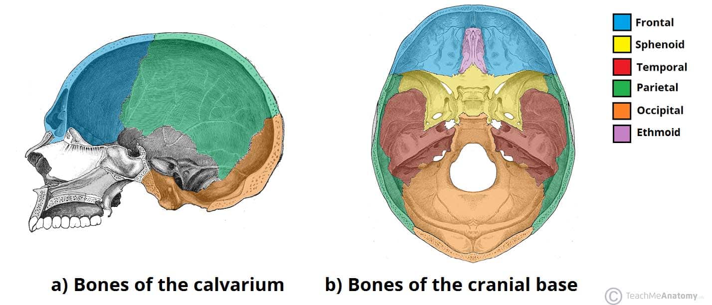


Bones Of The Skull Structure Fractures Teachmeanatomy
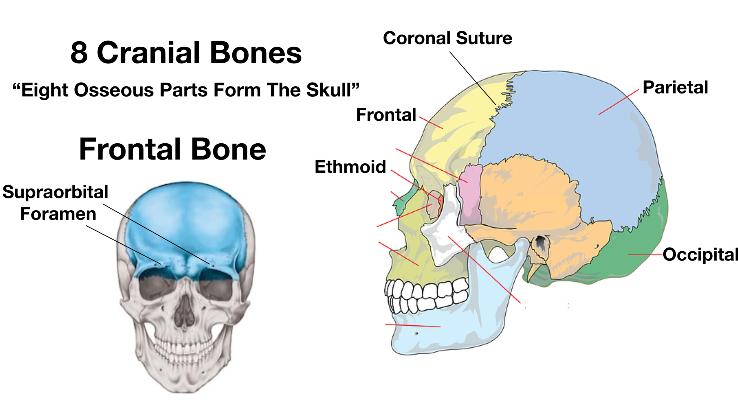


Skull Anatomy Cranial Bone And Suture Labeled Diagram Names Mnemonic Ezmed
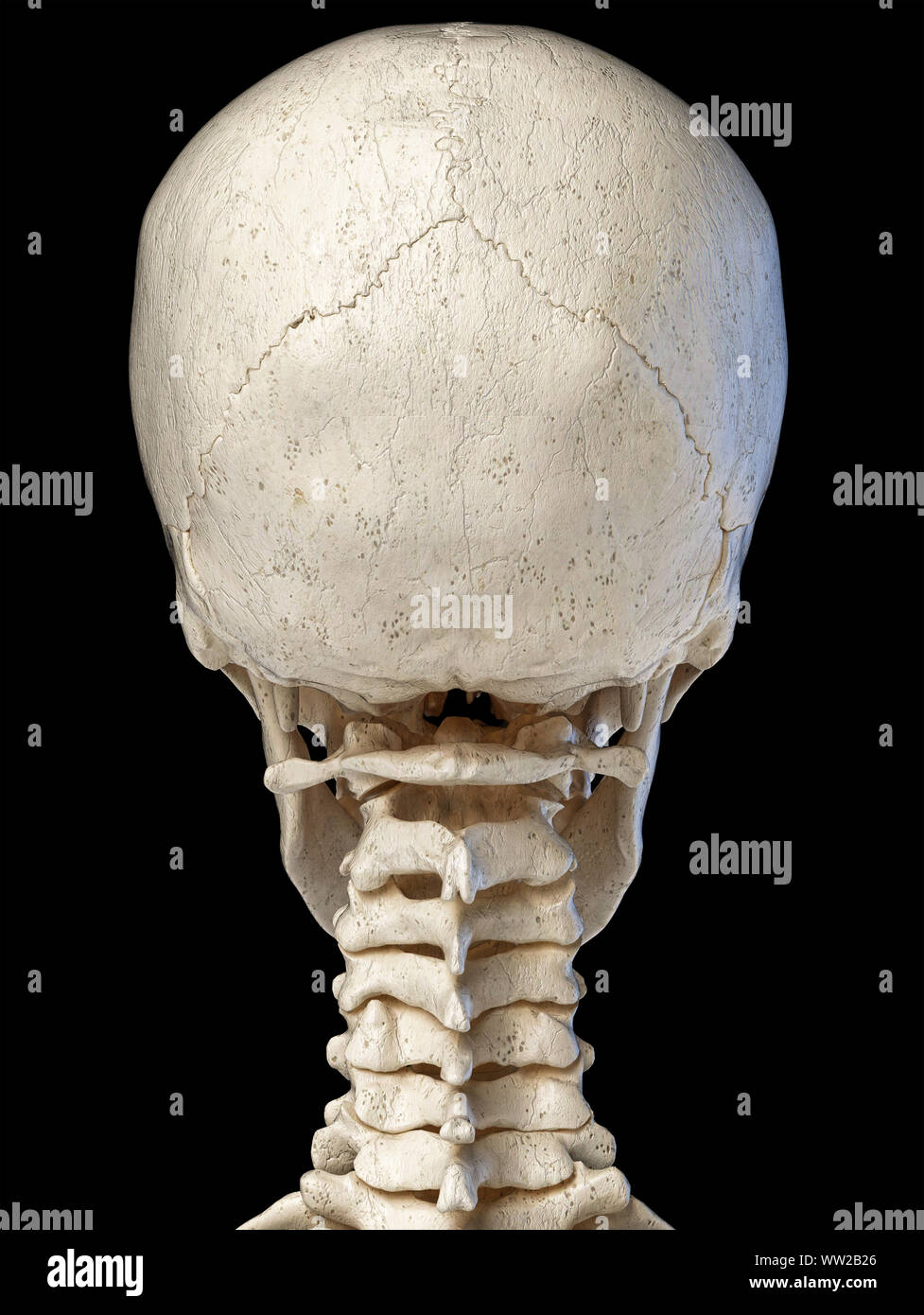


Human Head Skull Anatomy 3d Illustration Rear View On Black Background Stock Photo Alamy


Back Of Skull Bones Diagram Quizlet


The Skull Boundless Anatomy And Physiology
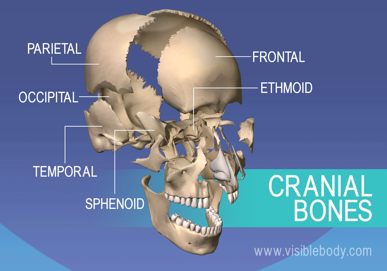


Axial Skeleton Learn Skeleton Anatomy
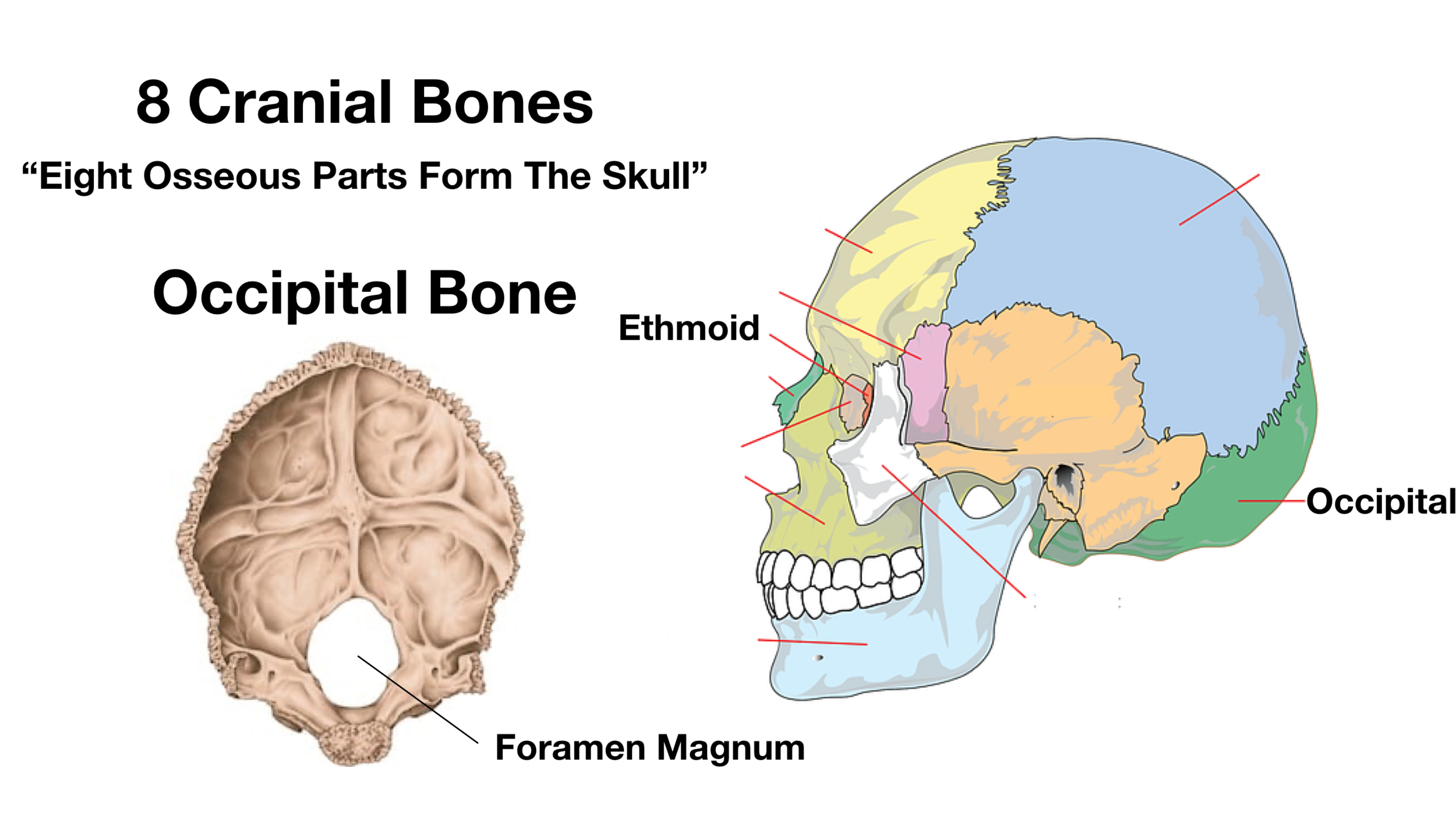


Skull Anatomy Cranial Bone And Suture Labeled Diagram Names Mnemonic Ezmed
:max_bytes(150000):strip_icc()/human-skull-with-veins-and-arteries--rear-view--1174640349-490cb7f8593945c4b1690b152e6a4074.jpg)


Occipital Artery Anatomy Function And Significance
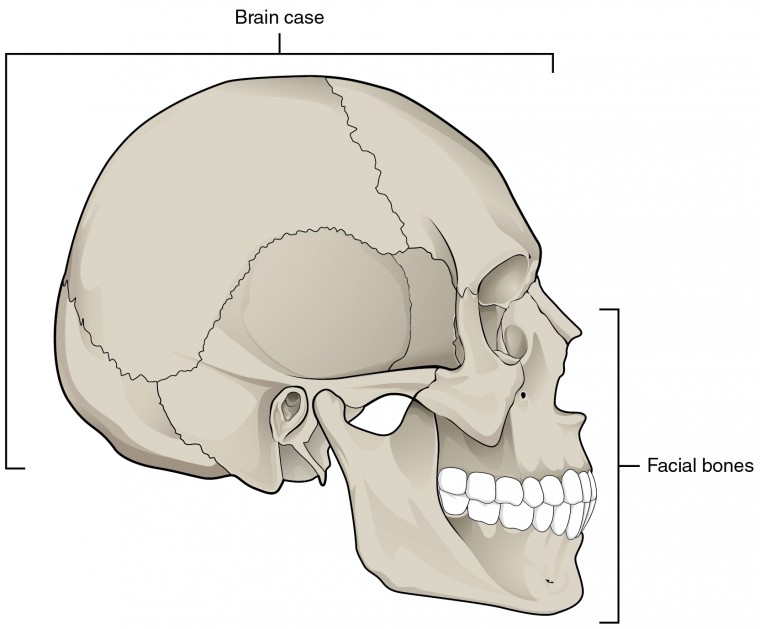


The Skull Anatomy And Physiology I
:background_color(FFFFFF):format(jpeg)/images/library/9490/skull-posterior-lateral-views_english.jpg)


Posterior And Lateral Views Of The Skull Anatomy Kenhub


1


Bones Of The Head Atlas Of Anatomy


Occipital Neuralgia And Suboccipital Headache C2 Neuralgia Treatments Without Nerve Block Or Surgery Caring Medical Florida
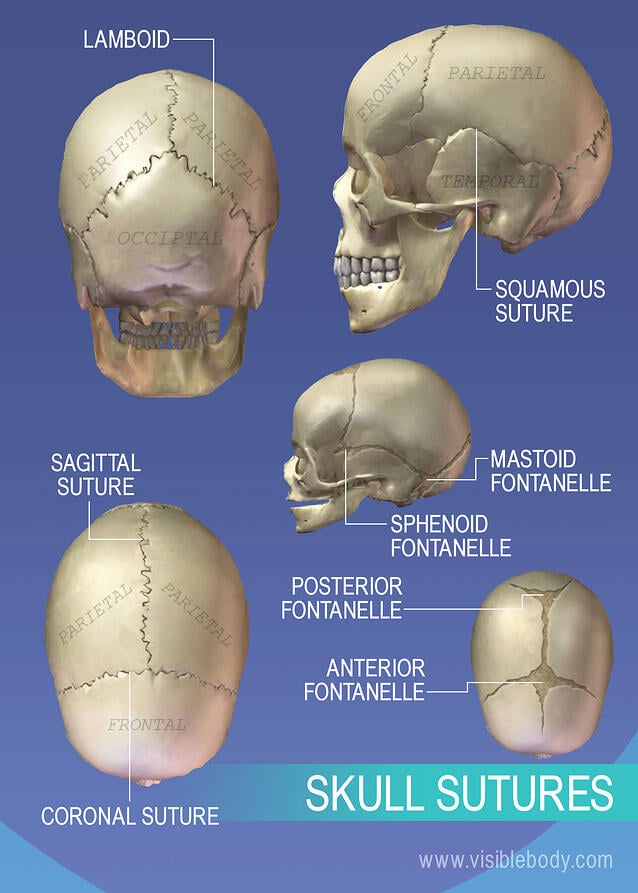


Axial Skeleton Learn Skeleton Anatomy
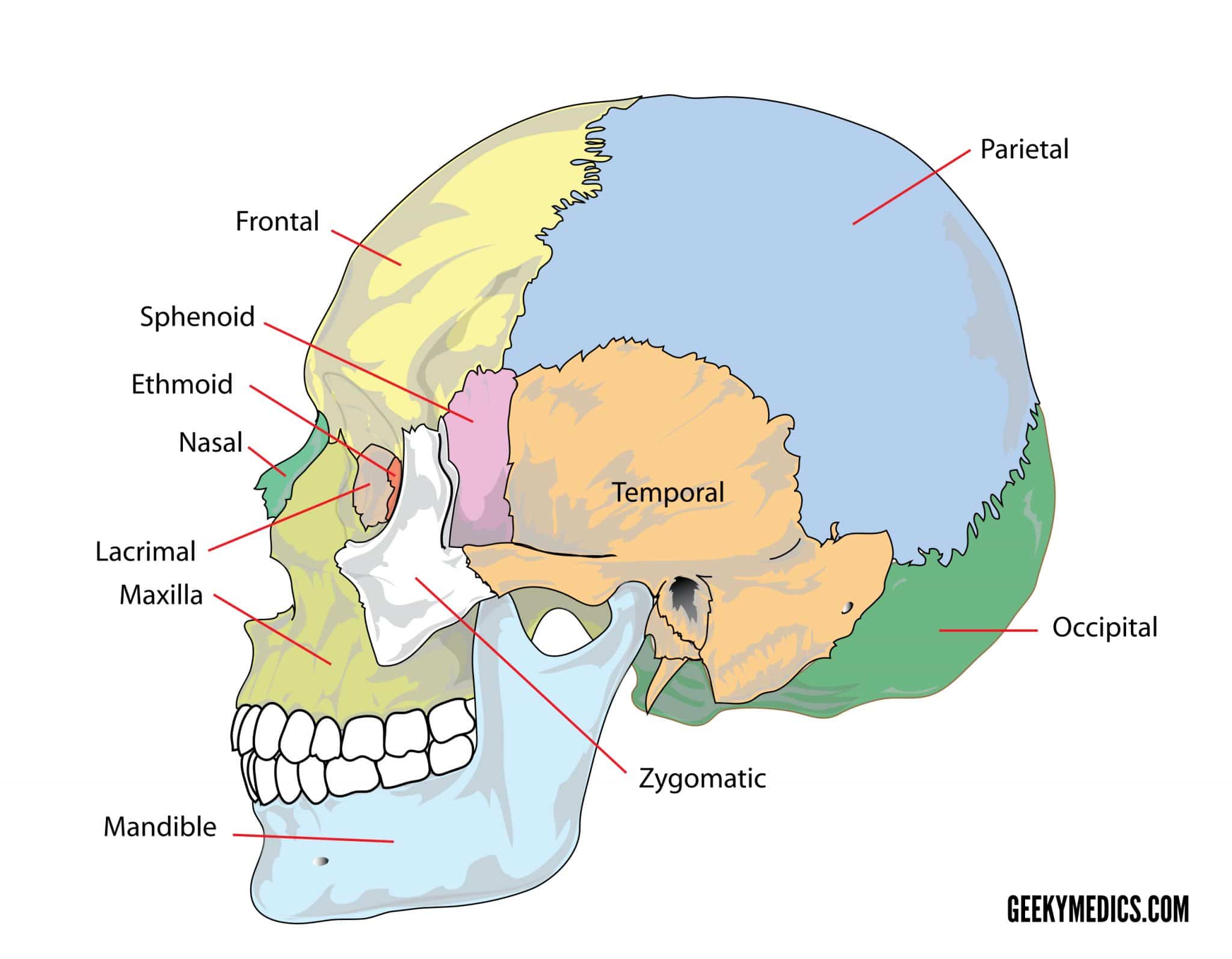


Bones Of The Skull Skull Osteology Anatomy Geeky Medics


Human Anatomy 3d Illustration Of Head With Skull Blood Vessels And Muscles On Black Background Rear View Stock Photo Picture And Royalty Free Image Image
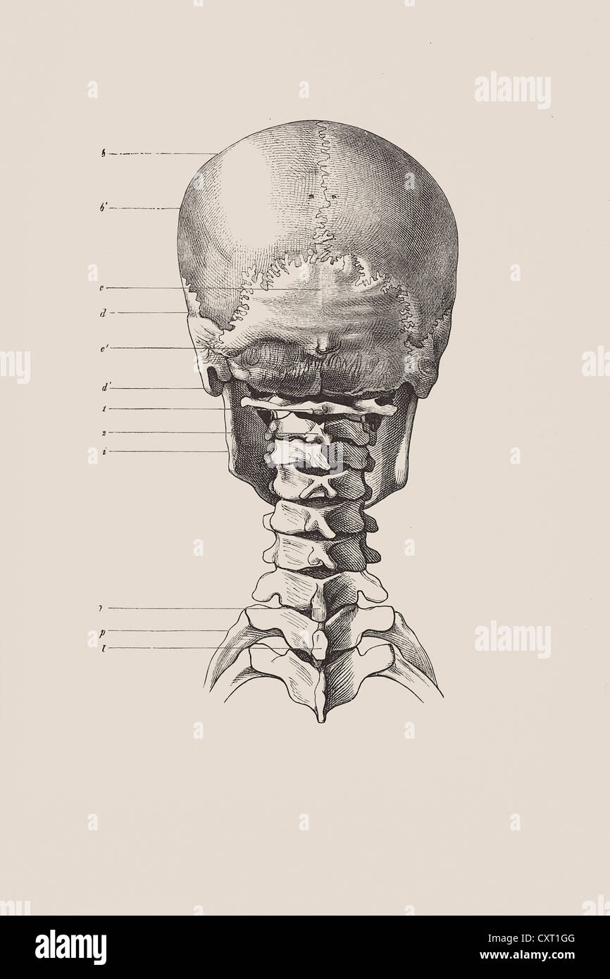


Cranial Skeleton From The Back Anatomical Illustration Stock Photo Alamy


Skeleton Anatomy 3d Atlas Human Anatomy Apps


Human Skull 3 4 Back View Skull Reference Human Skull Skull Anatomy


Bones Of The Skull Learn In 4 Minutes Youtube


Occipital Surgery For Flat Spots On Head Dr Barry L Eppley
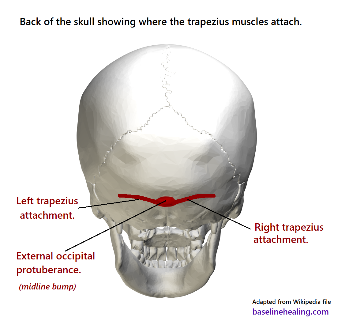


Upper Body To Base Line Connection The Trapezius Muscles
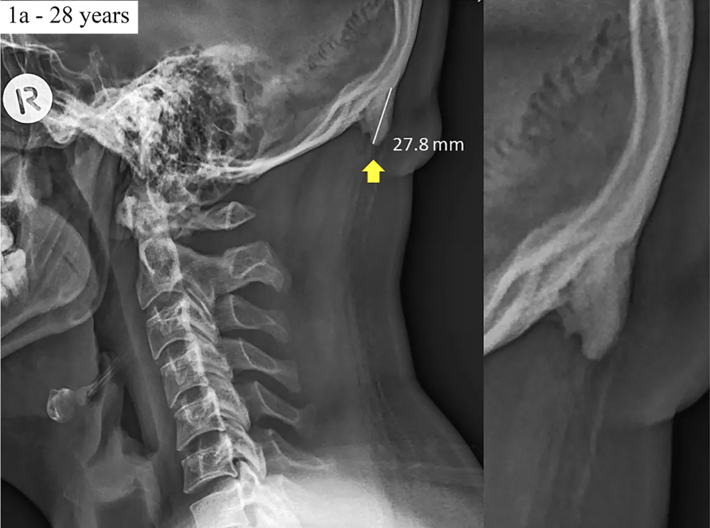


No Teens Aren T Growing Skull Horns Because Of Cellphones Time


The Skull Boundless Anatomy And Physiology


Cranial Sutures Anatomy Youtube


The Skull Anatomy And Physiology


Skull Anatomy Terminology Dr Barry L Eppley
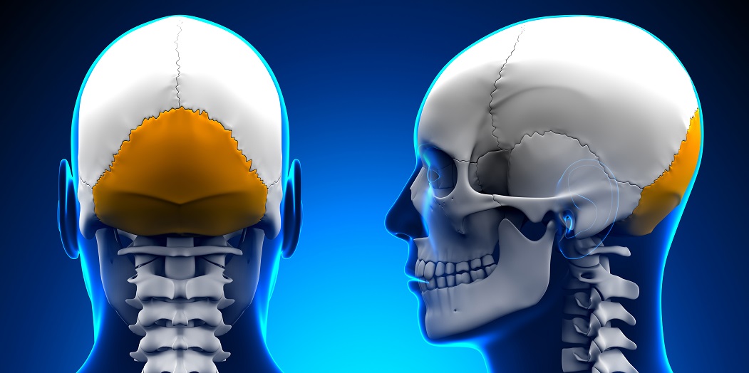


Occipital Bone The Definitive Guide Biology Dictionary


Cranial Bones Function And Anatomy Diagram Conditions Health Tips
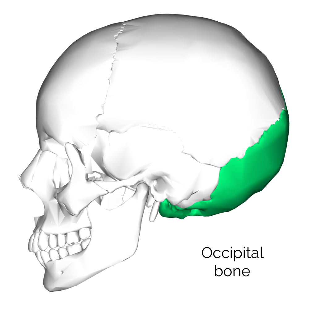


Bones Of The Skull Skull Osteology Anatomy Geeky Medics


Skull Scattered Back View Art Print Barewalls Posters Prints Bwc


Skull Anatomy Terminology Dr Barry L Eppley


Foramen Magnum Anatomy Britannica
:background_color(FFFFFF):format(jpeg)/images/library/10686/Posterior_view_of_the_skull.jpg)


Skull Anatomy Structure Bones Quizzes Kenhub


7 3 The Skull Anatomy Physiology
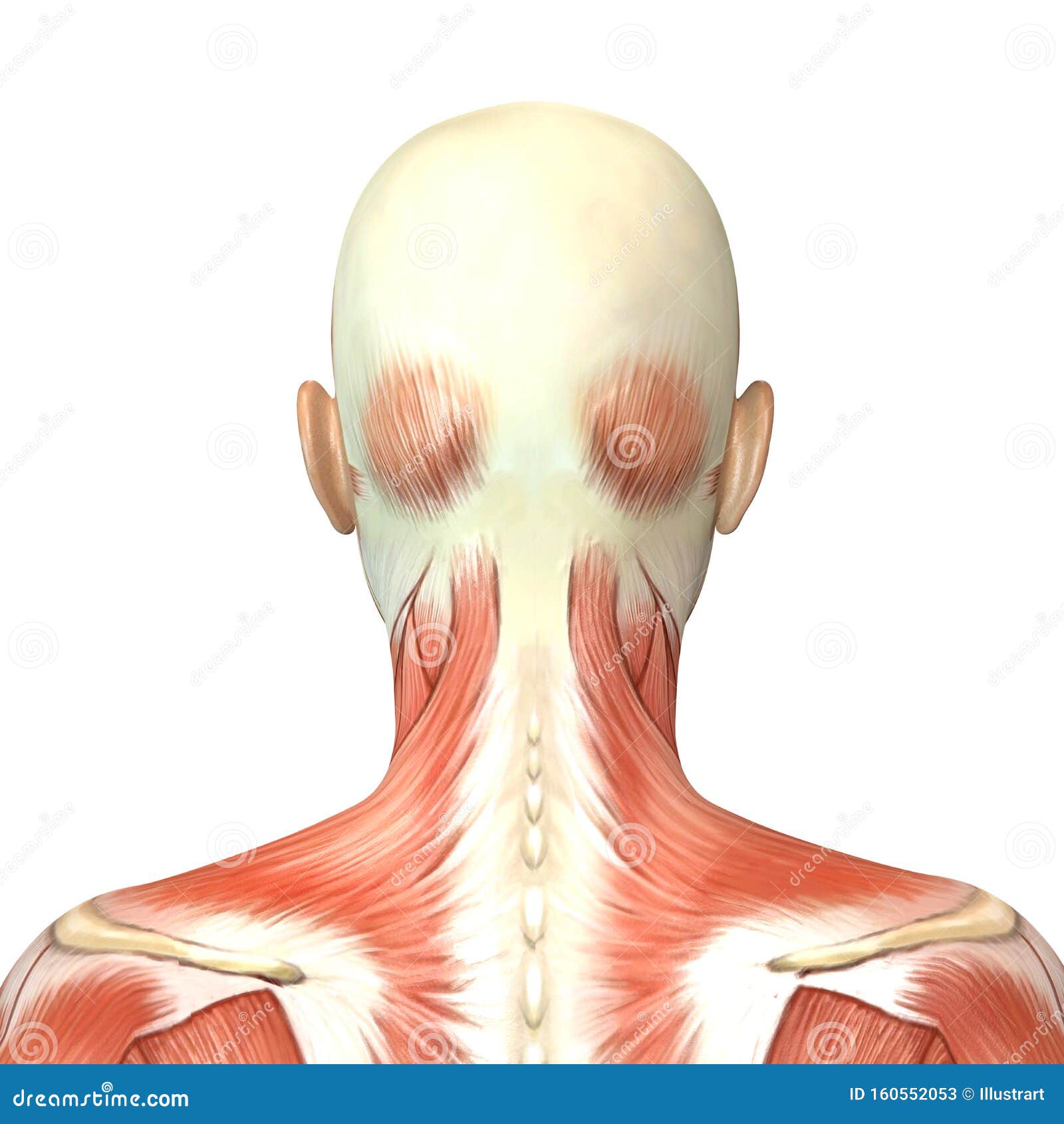


Back Head Anatomy Stock Illustrations 2 481 Back Head Anatomy Stock Illustrations Vectors Clipart Dreamstime
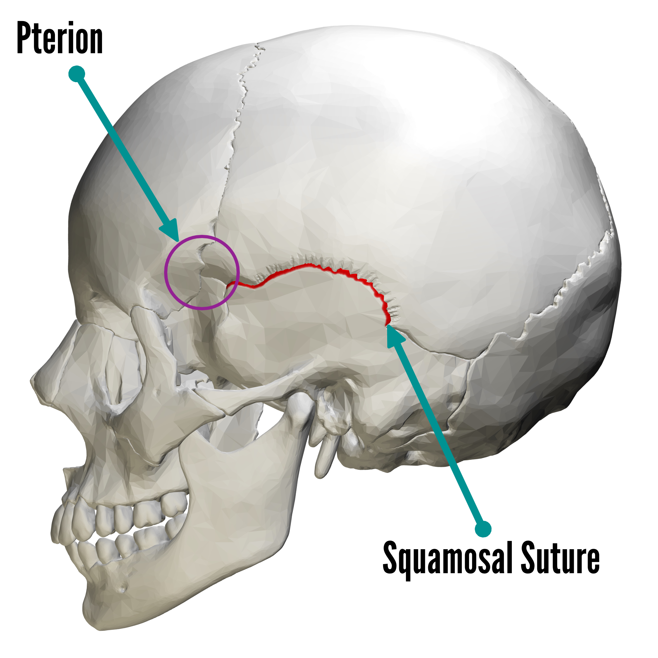


Bones Of The Skull Skull Osteology Anatomy Geeky Medics
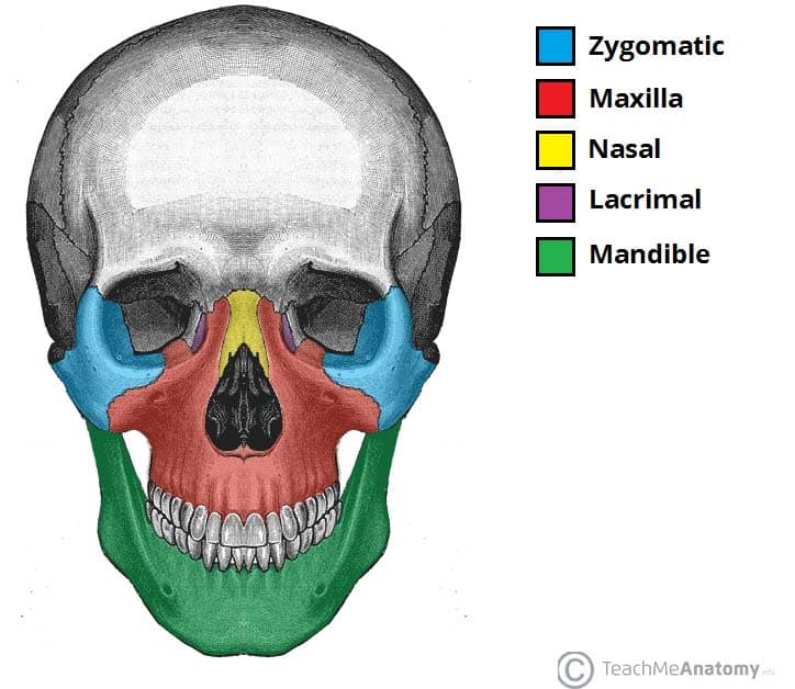


Bones Of The Skull Structure Fractures Teachmeanatomy
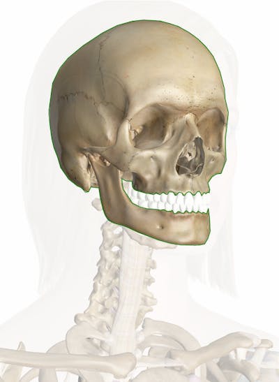


Skull Anatomy Pictures And Information


Upper Cervical Spine Disorders Anatomy Of The Head And Upper Neck
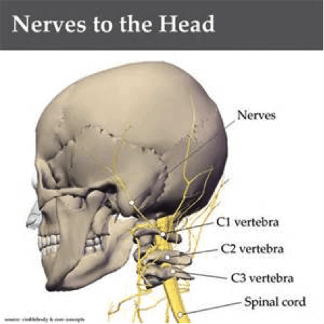


Neck Anatomy Pictures Bones Muscles Nerves



Posterior And Lateral Views Of The Skull Anatomy Kenhub


Skull Wikipedia


Skull Wikipedia
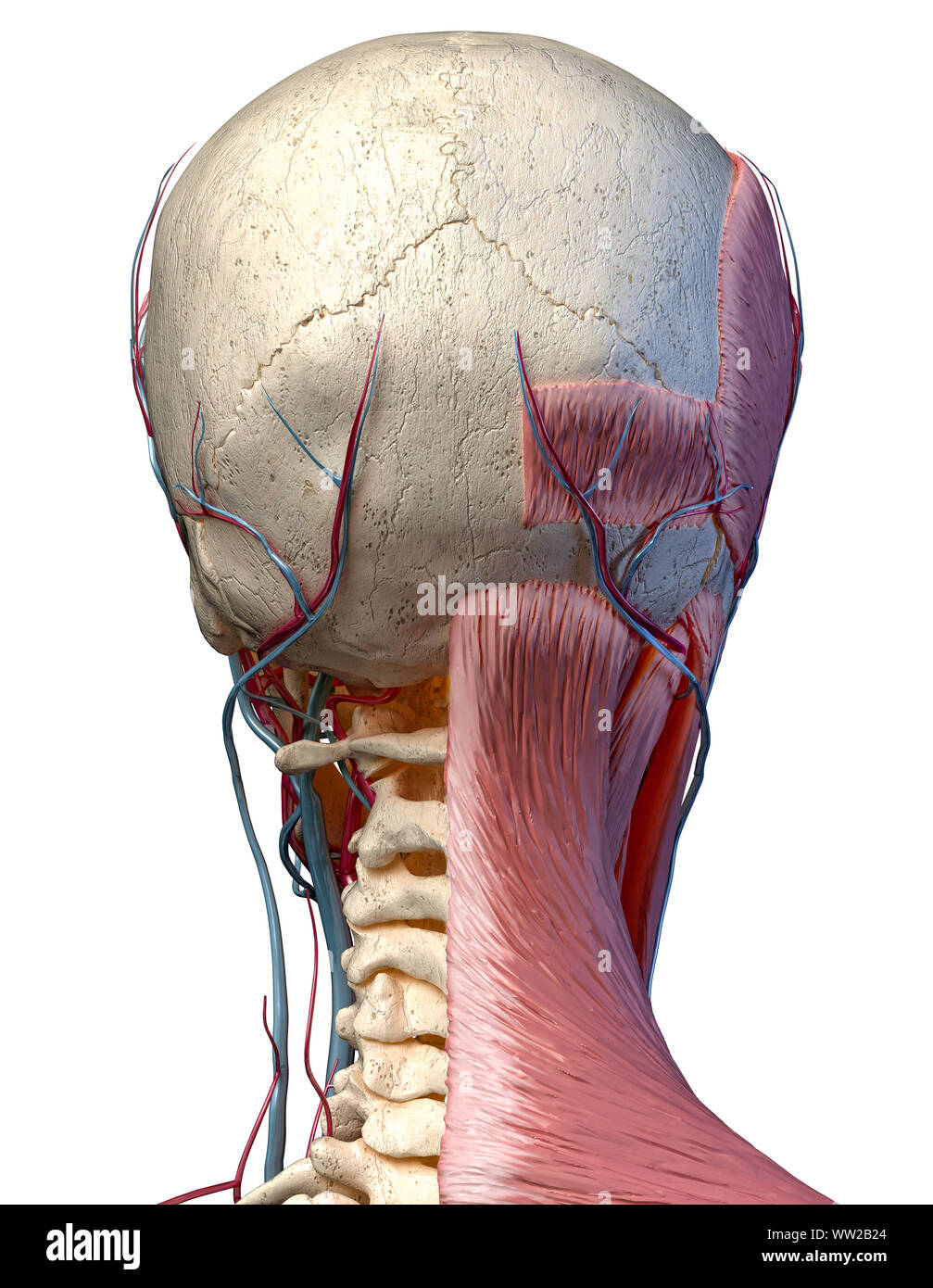


Human Anatomy 3d Illustration Of Head With Skull Blood Vessels And Muscles On White Background Rear View Stock Photo Alamy
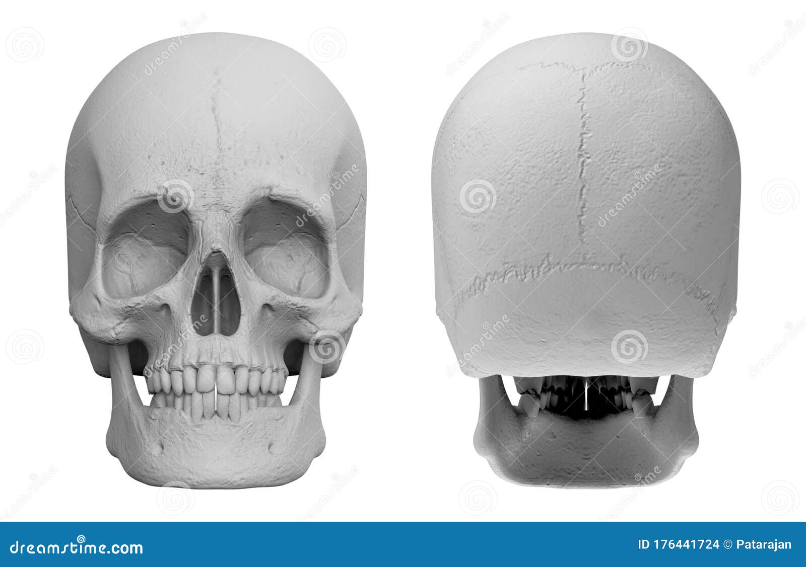


3d Rendering Set Of Front And Back Side Of Human Head Skull Bone Isolated On White Background Stock Illustration Illustration Of Human Background


Why Do I Have A Bone Bump On The Back Of My Skull Quora


Frontal Bone Human Skull


Occipital Surgery For Flat Spots On Head Dr Barry L Eppley


Skull Anatomy Terminology Dr Barry L Eppley
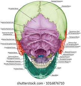


Skull Anatomy Images Stock Photos Vectors Shutterstock


Bones Of The Skull Structure Fractures Teachmeanatomy


7 3 The Skull Anatomy Physiology


Skull Definition Anatomy Function Britannica


Bones Of The Skull Skull Osteology Anatomy Geeky Medics


Temporal Bone Anatomical Diagram Function And Injuries


0 件のコメント:
コメントを投稿